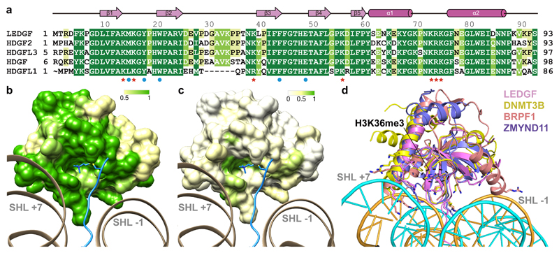
Figure 3. Conserved mode of nucleosome-PWWP interaction.
(a) Sequence alignment of PWWP domains within the HDGF-related protein (HRP) subfamily. Residues involved in H3K36me3 recognition are marked with blue circles; residues involved in DNA interaction are marked with red stars. Other conserved residues are highlighted from green to yellow with decreasing conservation. Secondary structure elements of the PWWP domain are shown above the sequences. (b) Conservation of PWWP domain surface within the HRP subfamily. Surface of the PWWP domain colored according to sequence conservation from green (identical) via yellow (conserved) to white (non-conserved). The H3 N-terminal tail is shown in blue. (c) Conservation of PWWP domain surface over all protein families. Color code as for (b). Note that both DNA patches and the methyllysine-binding region are at least partially conserved. (d) Superposition of known H3K36me3 peptide bound PWWP domain structures onto the nucleosome-PWWP structure presented here. PWWP domains of LEDGF, DNMT3B, BRPF1 and ZMYND11 are shown in cartoon and colored as indicated.

