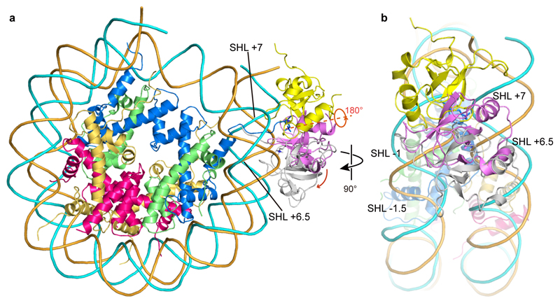
Extended Data Fig. 5. Comparison of the location of the PWWP domain in our nucleosome-PWWP complex structure with previously proposed models.
a. Front view of the comparison with two models proposed for LEDGF (gray) 25 or its highly conserved homolog HDGFL3 (yellow)26 with our structure (pink). Whereas in one model (yellow) the domain is rotated by around 180 degrees and shifted to SHL -1 on one DNA gyre, in another model (Gray) the domain is moved to SHL +6.5 and -1.5, and placed in the major groove of the DNA gyres.
b. Side view of the comparison shown in panel a.

