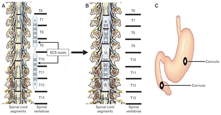
Figure 1.
Schematic figures of the location of the leads on the spinal cord and cannulae in the stomach and intestine in dogs after surgery. (A, B) Schematic figures of the exposed spinal cord to depict the location of leads alongside the spinal vertebrae (A: T7–T12) to cover the spinal cord segments (B: T8–T12). (C) Shows the locations of cannulae in both stomach and duodenum for inserting the catheter in the stomach to measure the motility index (MI) using high-resolution manometry as well as performing gastric emptying test through the duodenal cannula. SCS, spinal cord stimulation.

