Fig. 4.

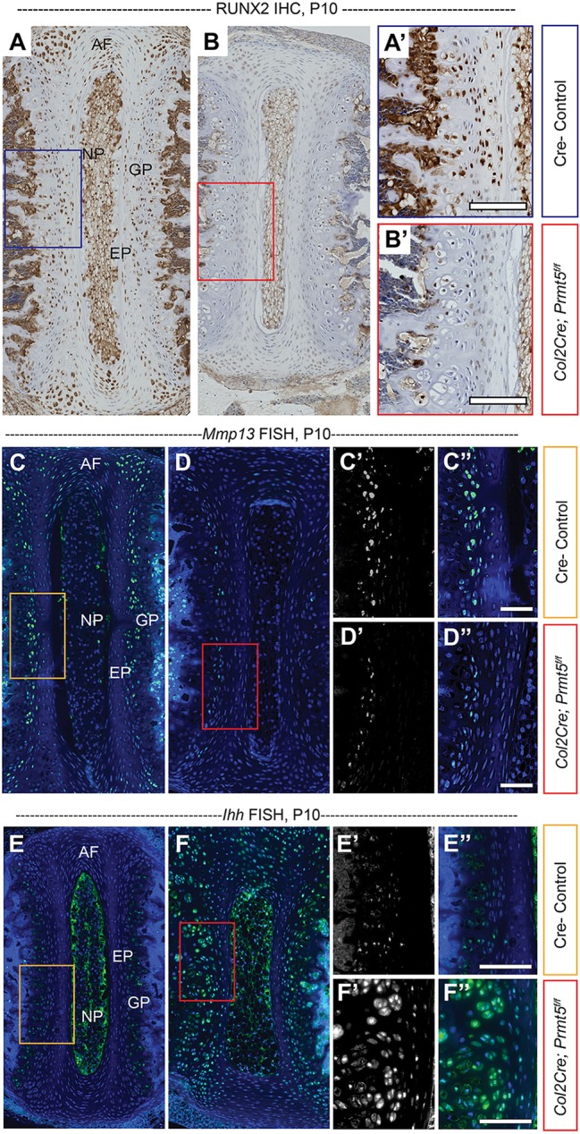
PRMT5 regulates the expression of RUNX2, Mmp13 and Ihh. (A,B) IHC analysis of RUNX2 in thoracic spine sections of Cre– control (A) or Col2Cre;Prmt5f/f mutant (B) mice at P10, demonstrating reduced RUNX2 expression in the endplate and growth plate of the mutant mice. The boxes outline the areas shown at higher magnification in A′ and B′. (C,D) Fluorescent in situ hybridization (FISH) analysis of Mmp13 in thoracic spine sections of Cre– control (C) or Col2Cre;Prmt5f/f mutant (D) mice at P10, with 4′,6-diamidino-2-phenylindole (DAPI; nuclei) in blue. Reduced Mmp13 signal was detected in the growth plate of the mutant mice. C′ and D′ are grayscale Mmp13 fluorescent in situ channels; C, C″, D and D″ are pseudocolored merged channels. The boxes outline the areas shown at higher magnification in C′, C″, D′ and D″. (E,F) FISH analysis of Ihh in thoracic spine sections of Cre– control (E) or Col2Cre;Prmt5f/f mutant (F) mice at P10. Induced expression of Ihh was detected in the growth plate of the mutant mice. E′ and F′ are grayscale Ihh fluorescent in situ channels; E, E″, F and F″ are pseudocolored merged channels. The boxes outline the areas shown at higher magnification in E′, E″, F′ and F″ (n=3 for each group). Scale bars: 100 µm. AF, annulus fibrosus; EP, endplate; GP, growth plate; NP, nucleus purposes.
