Fig. 4.

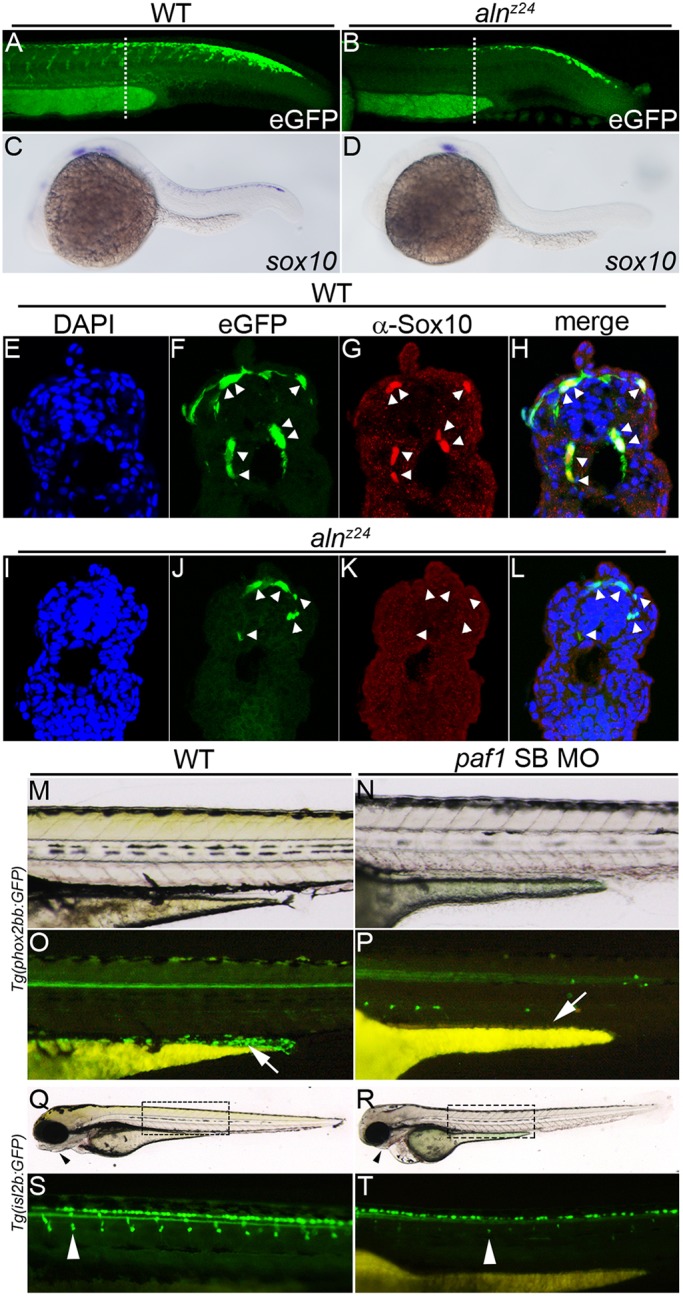
Paf1 is necessary to maintain NC gene expression, identity and the normal complement of multipotent premigratory NC. (A,B) Confocal images of eGFP expression in 26-28 hpf wild-type (A) and alnz24 mutant (B) embryos carrying the Tg(-4.9sox10:egfp)ba2 transgene. Dashed lines indicate approximate plane of section in E-L. (C,D) sox10 gene expression detected by whole-mount in situ hybridization in 24 hpf wild-type (C) and alnz24 mutant (D) embryos. (E-L) Transverse sections through the trunks of 26-28 hpf wild-type (E-H) and alnz24 mutant (I-L) transgenic embryos. In wild-type embryos, eGFP expression colocalizes with Sox10 protein expression (NC cells, indicated by arrowheads) (F-H). In contrast, all migrating GFP+ cells (arrowheads) in the trunk of alnz24 mutant embryos do not express Sox10 (J-L). (M-P) Lateral views with anterior towards the left of 72 hpf wild-type (M,O) and low dose paf1 MO-injected (N,P) Tg(phox2bb:GFP) embryos. GFP-expressing enteric neurons (arrow) are present in the gut of wild-type embryos (M) and are completely absent (arrow) in embryos with reduced paf1 function (P). GFP+ cells in paf1 MO-injected embryos are likely sympathetic neurons. (Q-T) Lateral views with anterior towards the left of 72 hpf wild-type (Q,S) and low dose paf1 MO-injected (R,T) Tg(isl2b:GFP) embryos. Reduction of paf1 function diminishes melanophore formation, and results in cardiac edema and loss of jaw structures (arrowheads) in MO-injected embryos (R) when compared with wild-type embryos (Q). GFP fluorescence marking DRG cells (arrowheads) in wild-type (S) and paf1 MO-injected embryos (T). DRG cell formation in paf1 MO-injected embryos is significantly reduced (T).
