Fig. 3.
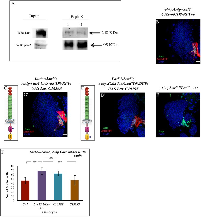
Lar physically and genetically interacts with InR. (A) pInR, immunoprecipitated from third instar larval lysate, shows an association with Lar, demonstrating a physical interaction between InR and Lar. (B-E) No rescue of niche cell number occurred upon overexpressing mutated PTPD1 (C1638S) domain in a Lar13.2/Lar5.5 background (C,C′), when compared with Lar13.2/Lar5.5 (E), whereas overexpressing mutated PTPD2 (C1929S) domain in Lar13.2/Lar5.5 (D,D′) resulted in a niche cell number (Antp, green) that was comparable with the control (B). (C,D) Schematic representation of the Lar protein (an RPTP), which has three Ig (immunoglobulin) domains (red circles), nine Fn-III (fibronectin type III) domains (green) and a transmembrane domain (gray); the pink indicates the PTP D1 (catalytic) domain and the yellow is the PTPD2 domain. The constructs used are mutated at PTPD1 (C1638S) and PTPD2 (C1929S) domains (denoted by an asterisk), respectively. (F) Quantification of the niche cell number in the above genotypes (n=9, P=8.458×10⁻⁶ for WT versus Lar13.2/Lar5.5, P=0.103 for Lar13.2/Lar5.5 versus UAS C1638S, P=0.0003 for Lar13.2/Lar5.5 versus UAS C1929S; two-tailed unpaired Student's t-test). The genotype of the larvae is described in the panels. The niche is marked with Antp antibody (green) in B,C′,D′,E and red represents Antp-Gal4.UASmcD8 RFP in B,C′,D′. Data are mean±s.d. ***P<0.0005. NS, not significant. Scale bars: 20 µm.

