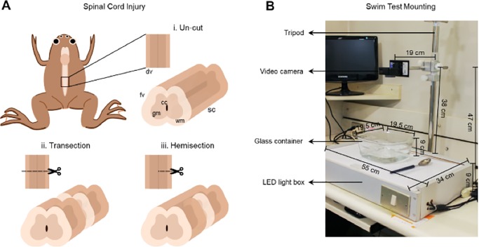
Fig. 6.
Experimental Setup. (A) Illustration of spinal cord injuries in the 6th vertebrae of X. laevis froglets in the dorsal and frontal views, (i) uninjured, (ii) transected and (iii) hemisected animals. (B) Picture of the setup used to record the movies of froglets. The tripod where the video camera is placed, a box internally illuminated with LED lights, a glass container with Barth solution and a spoon and a Pasteur pipette for swimming stimulation of froglets. fv, frontal view; dv, dorsal view of the spinal cord; sc, spinal cord; gm, grey matter; wm, white matter; cc, central canal.

