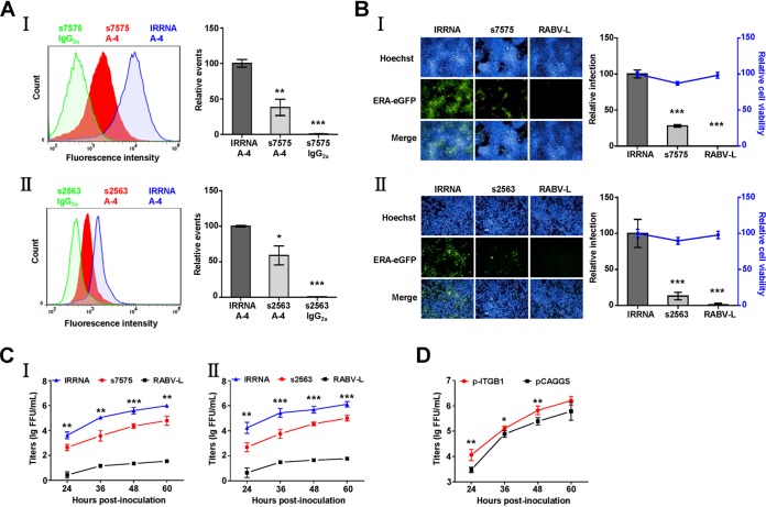
FIG 1.
siRNA silencing and overexpression of ITGB1 affect RABV infection in cells. (A) Transfection with siRNA s7575 or s2563 resulted in downregulation of ITGB1. HEK293 (I) or N2a (II) cells were transfected with siRNA s7575 or s2563, respectively, at 37°C for 48 h. Cells were collected and stained with mouse anti-ITGB1 MAb (A-4) and fluorescein isothiocyanate (FITC)-mouse IgG and analyzed using flow cytometry. siRNA s7575- or s2563-transfected cells stained with mouse IgG2a and the irrelative siRNA (IRRNA)-transfected cells stained with A-4 were used as controls. The amount of cell surface ITGB1 was shown by comparison with that of the IRRNA-transfected cells stained with A-4 group, which was set as 100. The statistical differences were assessed using Student's t test. *, P < 0.05; ***, P < 0.001. (B) Downregulation of ITGB1 inhibited ERA-eGFP infection. HEK293 (I) and N2a (II) cells were transfected with siRNAs at 37°C for 48 h and infected with ERA-eGFP at a multiplicity of infection (MOI) of 0.1 at 37°C for 48 h. siRNA targeting the RABV L gene (RABV-L) and IRRNA were used as controls. Cells were used to detect cell viability using a CellTiter-Glo kit and stained for imaging using the PerkinElmer Operetta high-content system, and infection rate of RABV was analyzed using Columbus software. Cell viability and infection rate were shown as the relative cell viability and relative infection compared with those of the IRRNA-transfected group, which were set as 100. (C) Downregulation of ITGB1 inhibited the growth of ERA-eGFP. HEK293 (I) and N2a (II) cells were transfected with siRNAs at 37°C for 48 h and infected with ERA-eGFP at an MOI of 0.1. The supernatants were harvested at different time points for virus titration, and virus titers were determined in BSR cells and expressed as focus-forming units (FFU) per milliliter. (D) Overexpression of ITGB1 enhanced growth of ERA-eGFP. HEK293 cells were transfected with plasmid expressing ITGB1 (p-ITGB1) at 37°C for 36 h and infected with ERA-eGFP at an MOI of 5 for 1 h at 4°C, washed with prechilled DMEM three times, and incubated with new DMEM (supplemented with 2% FBS) at 37°C. The plasmid vector (pCAGGS) was used as a control. Virus titers of the supernatants were determined as FFU/ml in BSR cells. All data were considered statistically significant using Student's t test. *, P < 0.05; **, P < 0.01; ***, P < 0.001.

