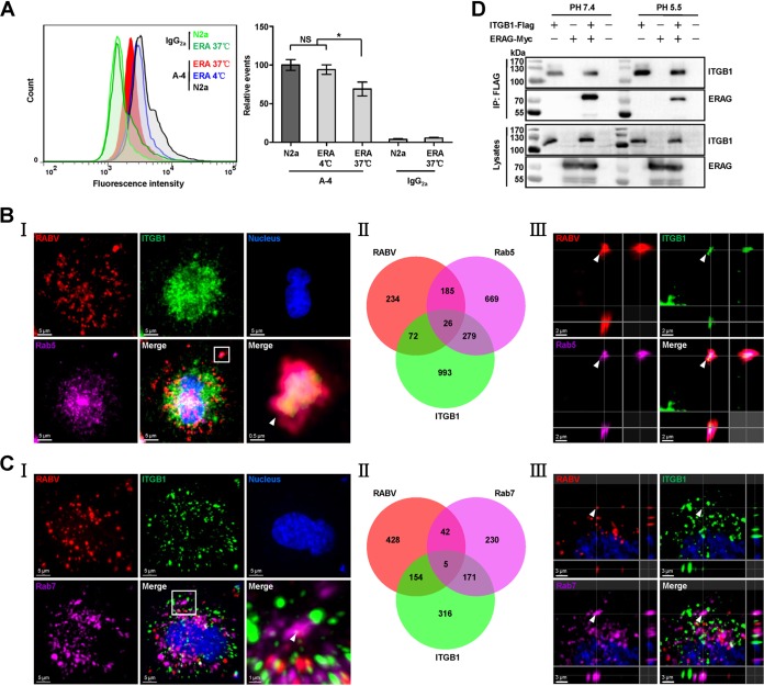
FIG 3.
RABV and ITGB1 are internalized into cells and transported to early and late endosomes together. (A) RABV infection results in downregulation of the cell surface ITGB1. N2a cells were infected with ERA at 37°C for 30 min, stained with A-4 and fluorescein isothiocyanate (FITC)-mouse IgG, and analyzed using flow cytometry. Noninfected cells (shown as N2a) stained with A-4 or mouse IgG2a, cells infected with ERA (shown as ERA) at 37°C and stained with mouse IgG2a, and cells infected with ERA at 4°C and stained with A-4 were used as controls. The amount of cell surface ITGB1 was shown as the relative events compared with that of the noninfected cells stained with A-4 group, which was set as 100. Significant differences were assessed using the Student's t test. NS, no significant difference. *, P < 0.05. (B and C) RABV and ITGB1 colocated in early and late endosomes. N2a cells were infected with recombinant RABV expressing an additional ERA nucleoprotein fused with a red fluorescent protein, mCherry (ERA-N/mCherry), at an MOI of 5 for 1 h at 4°C, washed with prechilled DMEM three times, and incubated with new DMEM (supplemented with 2% FBS) at 37°C for 30 min. Cells were stained using the tyramide signal amplification immunofluorescent method. RABV antigen (red), ITGB1 (green), Rab5 or Rab7 (purple), and the cell nuclei (blue) were examined in single-fluorescence channels. Colocalization of the RABV-ITGB1 complex with Rab5 or Rab7 was observed (I) and counted (II). The images in the lower right corner of panel I represent amplified random colocalization spots in the merged image within the small white box. (III) The 3D-rendered images were generated using Imaris software, and colocalization of the RABV-ITGB1 complex with Rab5 or Rab7 from the three-single fluorescence channels is indicated with the white arrowhead. (D) ITGB1-Flag interacted with ERAG-Myc under acidic conditions. ITGB1-Flag and ERAG-Myc plasmids were cotransfected in HEK293 cells at 37°C for 48 h, and ITGB1-Flag interacted with ERAG-Myc in co-IP assays with HEK293 cell lysates at pH 5.5 and pH 7.4.

