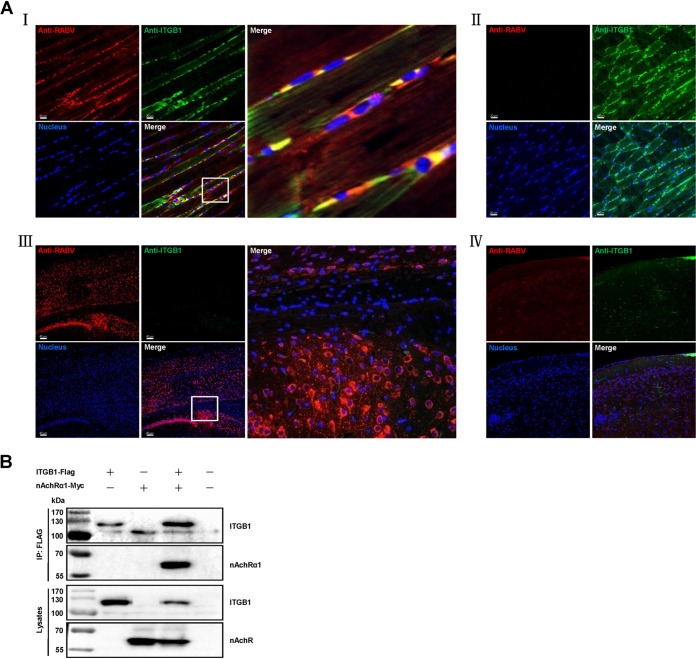
FIG 7.
RABV and ITGB1 coexist in RABV-inoculated muscle of mice and ITGB1 interacts with nAChRα1. (A) RABV street virus GX/09 and ITGB1 coexist in muscle cells but not cerebral cortex at the RABV-inoculated site. Six-week-old BALB/c mice were i.m. injected with 10 MLD50 GX/09 or i.c. injected with 5 MLD50 GX/09 for 4 days, and the RABV-inoculated thigh muscle and cerebral cortex were collected and fluorescently stained. Mice i.m. or i.c. injected with PBS were used as controls and processed in parallel. RABV antigen (red), ITGB1 (green), and the cell nuclei (blue) were observed in single-fluorescence channels using a Carl Zeiss LSM700 microscope. RABV antigen was observed in thigh muscle (I) and cerebral cortex (III). ITGB1 was stained in thigh muscle (I and II) but not cerebral cortex (III and IV) and colocalized with RABV in thigh muscle (I). The images on the right (I and III) represent amplified random spots in the merged image within the small white box. (B) ITGB1 interacted with nAChRα1. Plasmid ITGB1-Flag and nAChRα1-Myc were cotransfected in HEK293 cells at 37°C for 48 h, and ITGB1-Flag interacted with ERAG-Myc in co-IP assays with HEK293 cell lysates.

