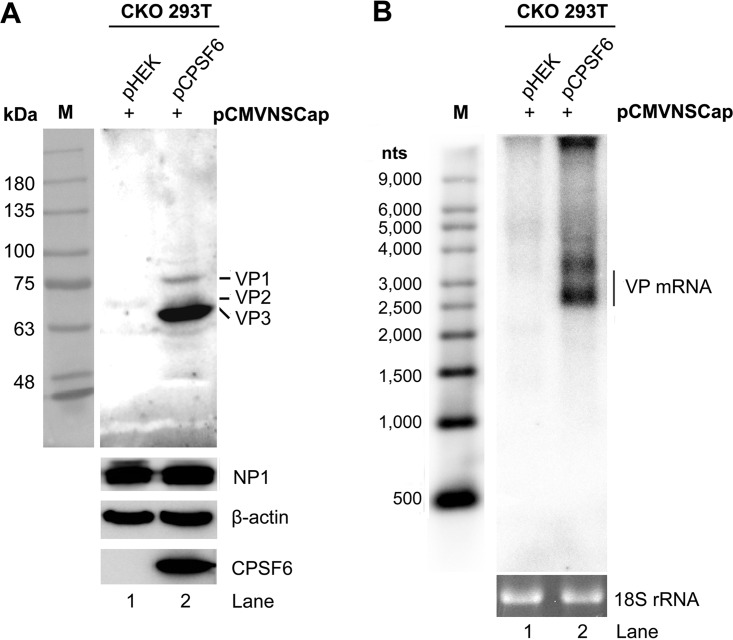
FIG 5.
Complementation of CPSF6 in CKO 293T cells rescues the expression of capsid proteins. CPSF6-knockout HEK293T cells (CKO 293T cells) were transfected with pCMVNSCap together with the pHEK empty vector or pCPSF6-Flag. Western blotting and Northern blotting were performed at 48 h posttransfection. (A) Western blot analysis of capsid proteins. Cell lysates were analyzed by Western blotting using anti-VP, anti-NP1, anti-CPSF6, and anti-β-actin antibodies. The detected protein bands are indicated. Lane M, protein size markers. (B) Northern blot analysis of VP-encoding mRNAs. Total RNAs were analyzed by Northern blotting using the Cap probe. Ethidium bromide-stained 18S rRNA bands are shown as the loading control, and the VP-encoded mRNA bands are indicated. A Millennium RNA ladder was used as a size marker (lane M).

