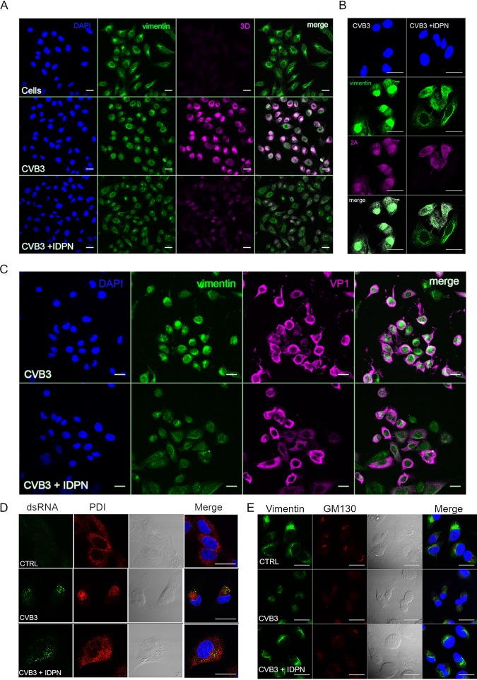
FIG 5.
Vimentin cage preferentially hosts nonstructural proteins. (A and B) Single sections showing location of 3D (magenta) (A) or 2A (magenta) (B) in the perinuclear area and vimentin (green) in control or CVB3-infected cells with or without IDPN treatment. Virus (4.43 × 10⁸ PFU/ml) was bound on ice for 1 h. After washing the excess virus away, infection was allowed to proceed for 5.5 h. IDPN was added after ice binding and left until the end of the experiment. (C) Single sections showing the location of VP1 diffusely in the cytoplasm in CVB3-infected cells with or without IDPN treatment. Infection was carried out as described for panel B. Scale bars, 20 μm. Representative images from at least three separate experiments are shown. (D and E) Single-section confocal images illustrating the effect of IDPN on ER (PDI) (5.5. h p.i) (D) and Golgi (GM130) (5.5 h p.i) (E). Scale bar, 20 μm. The images are representative of at least two separate experiments.

