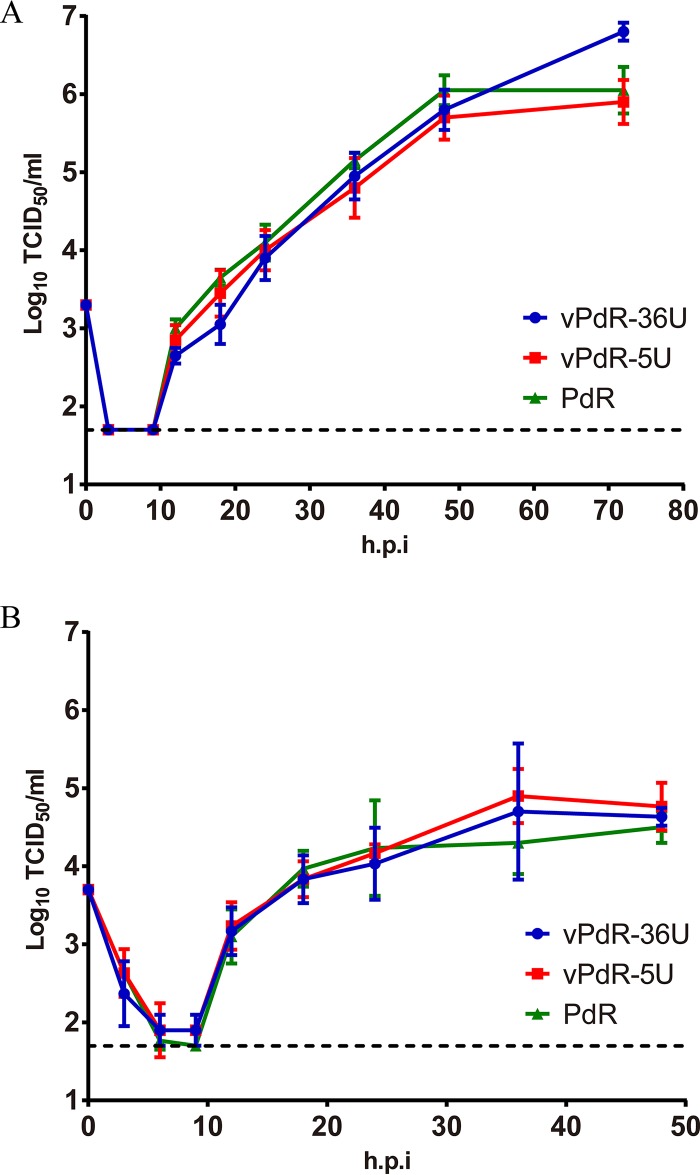
FIG 1.
Virus replication kinetics in PEDSV.15 cells and porcine macrophages. PEDSV.15 cells (A) and porcine monocyte-derived macrophages (B) were infected in quadruplicate and in triplicate, respectively, with CSFV PdR and with cDNA-derived vPdR-36U and vPdR-5U at an MOI of 0.02 TCID50/cell based on titers obtained in the homologous cell type (plotted on day 0). At the indicated hours postinfection (h.p.i), virus was harvested by one freeze-thaw cycle and the infectious titer was determined in PEDSV.15 (A) or in SK-6 cells (B) by endpoint dilution. The limit of detection of the titration (1.7 log10 TCID50/ml) is represented with a dashed line. Each point represents the mean titer from four parallel infections in panel A and three in panel B, with error bars showing the SDs.

