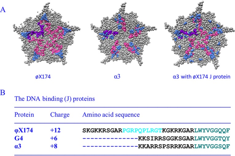
FIG 1.

Characteristics of microvirus J proteins and their structural influences on packaged DNA. (A) Fivefold axes of symmetry of ϕX174 filled with its own J protein (PDB 2BPA), α3 filled with its own J protein (PDB 1M06), and α3 filled with the ϕX174 J protein (PDB 1RB8). The coat protein is depicted in gray, and ordered DNA is shown in light blue. The leftmost J protein is depicted in purple, the remaining five are depicted in magenta (B) ϕX174, G4, and α3 J proteins. The conserved, coat protein-interacting, C-terminal amino acids are depicted in teal. The basic DNA-binding regions are depicted in black. The unique proline-rich region in the ϕX174 protein is depicted in cyan.
