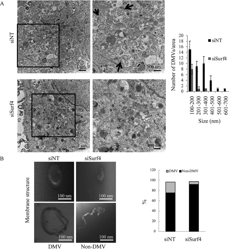
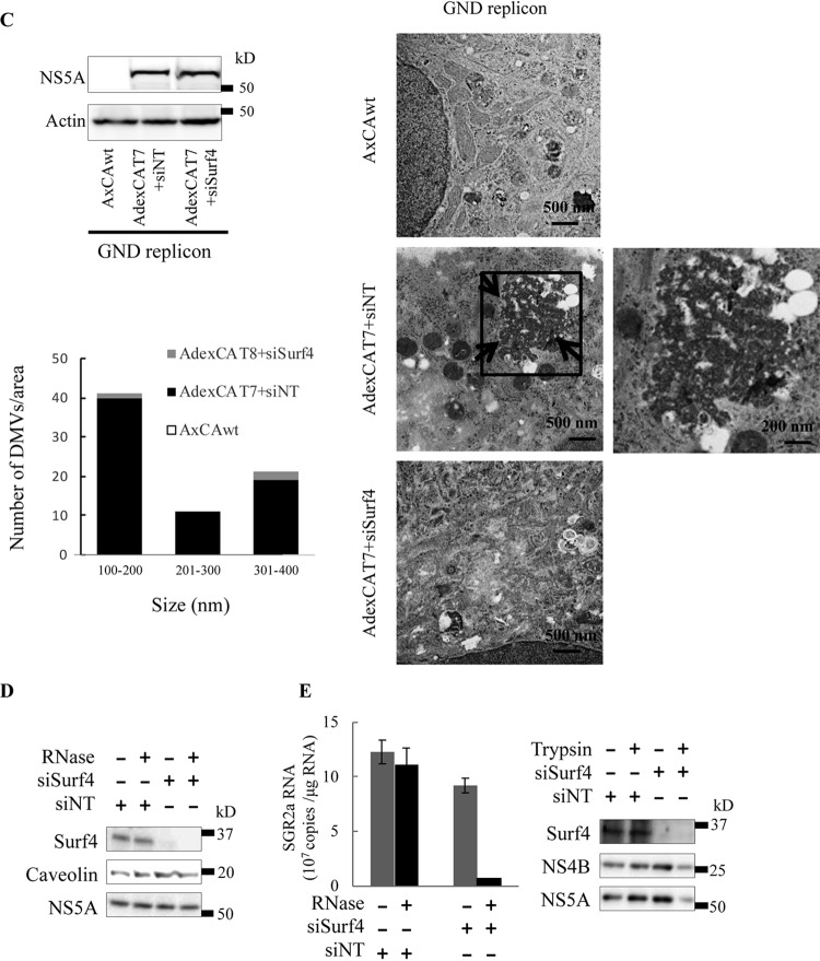
FIG 5.
Surf4 is involved in the formation of DMVs and structural integrity of HCV RNA replication complexes. (A) SGR2a cells were treated with 25 nM siSurf4 or siNT. At 3 days posttransfection, cells were examined by TEM (left). The right panels are magnified views of the black squares in the left panels. Arrows indicate HCV protein-induced membrane vesicles. The size and number of DMVs in the boxed areas of siRNA-treated cells in panel A were quantified (right). Values were obtained from three samples and are means plus SDs. (B) SGR2a cells were treated with siSurf4 or siNT for 3 days. Membrane fractions from treated cells were examined by TEM. Representative membranous structures are shown (left). The number of membrane structures in 10 randomly chosen squares (3,600 μm2/square) was determined. The percentage of DMV and non-DMV in siSurf4-treated or control cells was determined for more than 200 membrane structures (right). (C) Huh7 cells were infected with AdexCAT7, a recombinant adenovirus containing the bacteriophage T7 RNA polymerase, or AxCAwt, a control virus, and transfected with pSGRlucneoGND. The cells were then transfected with 25 nM siSurf4 or siNT. At 3 days posttransfection, cells were examined by immunoblotting and TEM. Arrows indicate HCV protein-induced membrane vesicles. The micrographs present low-magnification overviews; the boxed areas are enlarged in the corresponding panels on the right. The size and number of DMVs in the areas in left panel were quantified. Values were obtained from three samples and are means ± SDs. (D and E) SGR2a cells were treated with siSurf4 or siNT. Three days later, DRM from treated cells were digested with RNase A (500 ng/ml) at 37°C for 5 min as indicated, and then used to determine the HCV RNA levels by real-time RT-PCR (graph in panel E) and protein levels by immunoblotting using anti-Surf4, anti-NS5A, and anti-caveolin antibodies (D). SGR2a cells were treated with siSurf4 or siNT. Three days later, DRM from transfected cells were treated with trypsin (25 μg/ml) at 37°C for 10 min as indicated and then used to determine protein levels by immunoblotting using anti-Surf4, anti-NS4B, and anti-NS5A antibodies (blots in panel E). For real-time RT-PCR, data are averages of triplicate values with error bars representing SDs.


