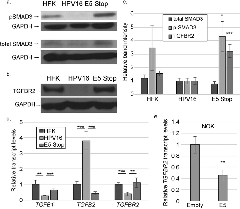
FIG 10.

TGF-β signaling is increased in E5 Stop cells. (a and b) Western blots showing the levels of SMAD3/phosphorylated SMAD3 (a) and TGFBR2 (b). (c) Quantification of results of immunoblotting experiments. (d) mRNAs for TGFB1, TGFB2, and TGFBR2 in HFK, HPV16+, and E5 Stop cells were measured by RT-qPCR. (e) Levels of TGFBR2 mRNA in hTert-immortalized NOKs stably transduced with an empty vector or a vector expressing 16E5 were measured using RT-qPCR. The blots shown are representative. *, P < 0.05; **, P < 0.01; ***, P < 0.001. The error bars indicate standard error of the mean.
