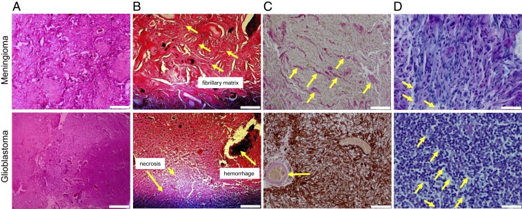
Fig. 2.
Histological images of 2 representative cases of GBM and MEN with demarcation of structures that potentially influence macroscopic viscoelastic properties. (A and B) H&E stains demonstrate fibrillary structures in MEN and necrotic regions in GBM (arrows). (Scale bar: A, 400 μm; B, 200 μm.) (C) Collagen is further revealed by Elastica van Gieson staining as light red/pink fibrils throughout the ECM in MEN and in the vascular and perivascular spaces in GBM (arrows), while the glial matrix is highlighted with Glial Fibrillary Acidic Protein (GFAP) immunostain. (Scale bar: 100 μm.) (D) Alcian blue staining highlights sulfated GAGs as light blue regions (arrow), showing abundance of GAGs in GBM and lower GAG density in MEN. (Scale bar: 40 μm.)

