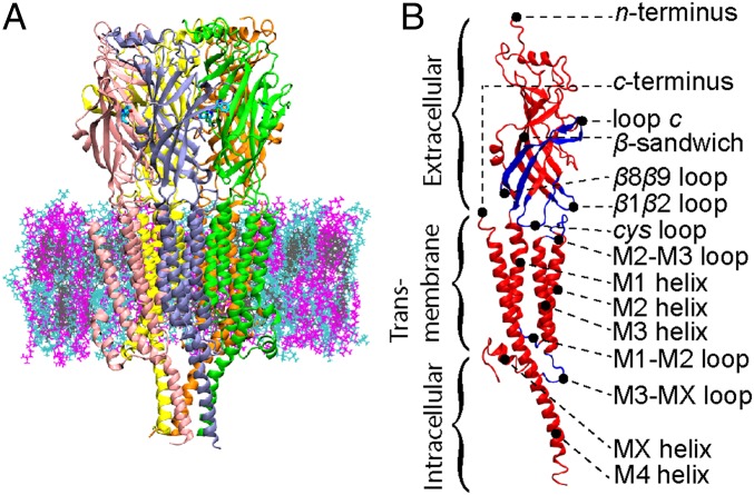
Fig. 1.
(A) Cross-section of 5-hydroxytryptamine 3A (5-HT3A) in a lipid membrane with 5-HT3A represented by secondary structure and colored by monomer (A, green; B, purple; C, pink; D, yellow; E, orange), lipids represented as lines, and lipid type represented by color: 1-palmitoyl-2-oleoyl-SN-glycero-3-phosphocholine (POPC), cyan; 1-stearoyl-2-docosahexaenoyl-sn-glyerco-3-phosphocholine (SDPC), magenta; cholesterol, gray; and 5-HT also represented as lines (aqua). (B) A single monomer of 5-HT3A represented by secondary structure and colored as red and blue to create contrast for specific secondary structure motifs.

