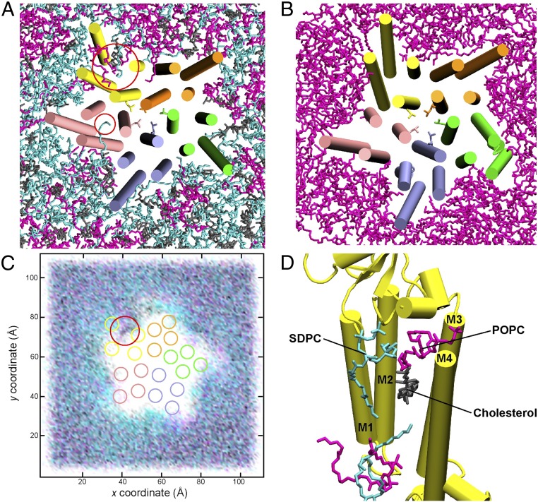
Fig. 5.
(A) Representative snapshot of 5-HT3A-5mM colored by monomer (A, green; B, purple; C, pink; D, yellow; E, orange) depicting transient penetration of the TMD by 1-stearoyl-2-docosahexaenoyl-sn-glyerco-3-phosphocholine (SDPC) (cyan) and sustained penetration by 1-palmitoyl-2-oleoyl-SN-glycero-3-phosphocholine (POPC) (magenta), SDPC, and cholesterol (gray). (B) Representative snapshot of 5-HT3A-5mM-POPC depicting no such penetration when only POPC is present. (C) Lipid density over 15 μs for 5-HT3A-5mM depicting clustering of SDPC around the TMD and sustained penetration of the TMD. (D) Snapshot of 5-HT3A-5mM monomer D (yellow) shown as secondary structure with penetrating lipids shown as sticks and other lipids removed for clarity.

