Fig. 1.
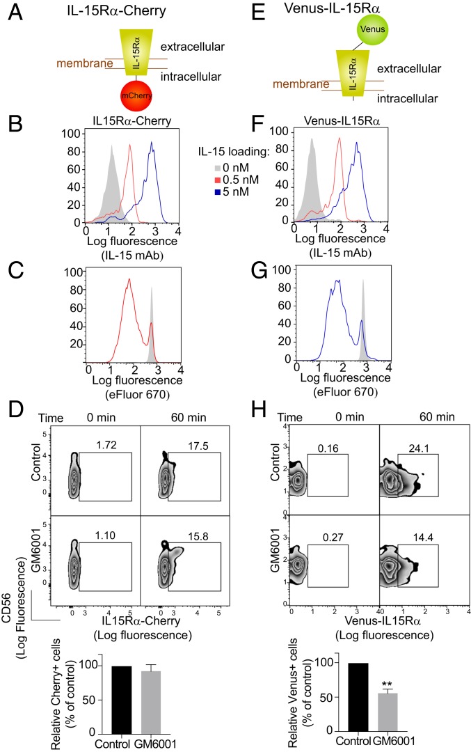
Transfer of membrane-associated IL-15Rα from trans-presenting cells to NK cells. (A) Schematic of the IL-15Rα-mCherry construct. (B) 221–IL-15Rα-mCherry cells were loaded with the indicated concentrations of IL-15 for 20 min, washed, and stained with an antibody to IL-15 and a secondary antibody coupled to PE. The control histogram (shaded) represents staining of cells not loaded with IL-15. (C) Proliferation of NK cells labeled with eFluor 670 and incubated with IL-15–loaded 221–IL-15Rα-mCherry cells (red) for 5 d. The shaded histogram represents NK cells incubated with 221–IL-15Rα-mCherry cells not loaded with IL-15. (D) 221–IL-15Rα-Cherry cells were preloaded with 5 nM IL-15 and incubated with primary human NK cells in the absence or presence of 0.4 μg/mL GM6001. Transfer of IL-15Rα-mCherry to NK cells was measured by flow cytometry. A representative experiment is shown. Bar graphs show the mean and SEM of 3 independent experiments relative to the control, untreated cells. (E) Schematic of the Venus-IL-15Rα construct. (F) 221–Venus-IL-15Rα cells treated as in B. (G) Proliferation of NK cells labeled with eFluor 670 and incubated with 221–Venus-IL-15Rα cells as in C. (H) 221–Venus-IL-15Rα cells were treated as in D. Transfer of Venus-IL-15Rα to the NK cells was measured by flow cytometry. Statistical analysis was performed using a 2-tailed t test. **P < 0.01.

