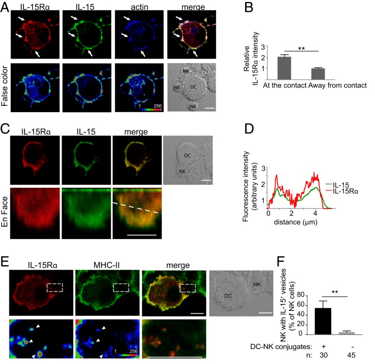
Fig. 2.
IL-15Rα is concentrated at DC–NK cell synapses and internalized into NK cells. (A) Human monocyte-derived DCs transfected with IL-15Rα-mCherry for 24 h were mixed with autologous primary human NK cells for 30 min. Conjugates were fixed, permeabilized, and stained with anti–IL-15 antibody and phalloidin. (Lower) False-color images. Arrows point to sites of IL-15Rα enrichment. (B) Enrichment of IL-15Rα at DC–NK cell synapses is represented as the fluorescence intensity at the contact site divided by the fluorescence intensity of a region in which there is no DC–NK cell contact. (C) Reconstruction of the synapse area; an en face view is shown. (D) Scan of fluorescence intensity of IL-15 and IL-15Rα along the dotted line in C. (E) DCs transiently transfected with IL-15Rα-mCherry were conjugated with NK cells for 1 h. IL-15Rα was detected in vesicles inside the NK cells. Some of those vesicles also contained MHC-II (arrowheads). (Lower) Magnification of the boxed area. (F) Frequency of NK cells in conjugates that had IL-15Rα–containing vesicles. n, number of cells per condition. The graph represents the mean ± SD of 3 independent experiments. Statistical analysis was performed using a 2-tailed t test. (Scale bar: 5 μm.) **P < 0.01.

