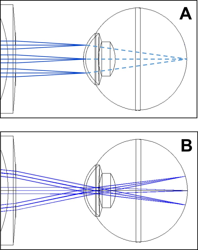
FIGURE 1. Differences in OCT scanning between anterior segment (A) and retina (B).
In each image, there are three example paths in solid blue representing a middle and two edge A scans. For anterior segment scanning (A), the scan proceeds from side to side (up and down in this image) and is focused on the anterior segment. Continuing on (illustrated as dashed light blue lines), these paths then converge on the same spot at the retina. Because the light for each A scan was focused at the anterior segment, they will be defocused at the retina. If recorded, the resulting retinal image would be a blurry image on one local spot at the retina, usually the foveal region. In regular retinal OCT scanning (B), The paths converge at the pupil and then fan out to scan the retina from side to side. The optics are also designed to focus light at the retina. These differences between the two systems make it not straight-forward to image both the anterior segment and the retina well in a single scan.

