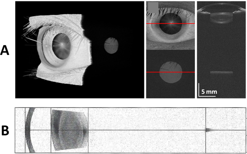
FIGURE 4. Images from long imaging depth OCT scanners.
A. The top row is from a research VCSEL based system with sufficient coherence length to image from the front to back of the eye.23 B. The bottom row is from a commercial swept-source OCT system (Zeiss IOLMaster 700). This more conventional swept-source system is designed to have a long imaging depth which allows it to image the cornea, phakic lens, and retina (small darker grey region on right of image in row B) in one single image.
Notice that in both these long imaging depth systems, the image of the retina (small grey disc in row A, small grey area on right in row B) is localized to the fovea and is laterally limited compared to a conventional retinal OCT image. The reason for this is due to the anterior segment scanning geometry (example in Fig. 1A) used by this these systems which limits imaging on the retina.

