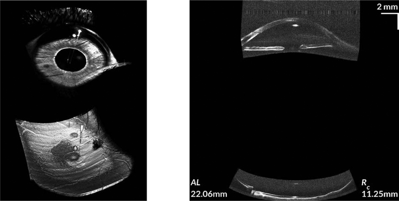
FIGURE 5. Simultaneous whole OCT of both the anterior segment and retina.
The image on the left is a volumetric rendering of the simultaneously acquired anterior segment and retina. From the volumetric rendering, the large fields of view on both the anterior segment and retina can be readily appreciated. The image on the right shows spatially corrected, simultaneously acquired cross-sections of the anterior segment and retina. Biometric information from the whole eye scan was used to perform the scan geometry and refraction corrections.28 (The left and right images are from two separate subjects. Additional detail on this whole eye OCT system can be found in McNabb, et al.27).

