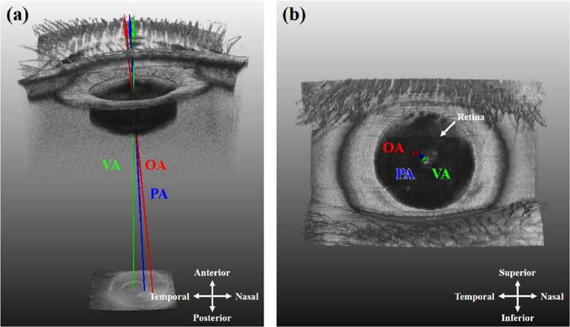
FIGURE 6. Using a whole eye OCT system to investigate axes of the eye.26.
Because a whole eye OCT system captures both the anterior segment (cornea apex, pupil) and the retina (fovea) simultaneously, the optical axis (OA), visual axis (VA), and pupillary axis (PA) can all be determined from the scan information. This enables investigation of relationships between these various axes.

