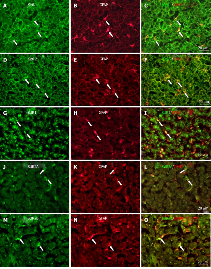
Figure 10.
Immunofluorescence double staining. Immunofluorescence double staining for Kir6.1, Kir6.2, SUR1, SUR2A, and/or SUR2B (A, D, G, J, and M) and GFAP [B, E, H, K, and N, marker of hepatic stellate cells (HSCs)] in no-fixed liver sections; Most of the GFAP+ cells (HSCs) colocalized with Kir6.1 (C, arrows) and/or Kir6.2 (F, arrows); There were few GFAP+ cells (HSCs) colocalized with SUR1 (i, arrows), SUR2A (L, arrows), and/or SUR2B (O, arrows). Bars: 20 µm.

