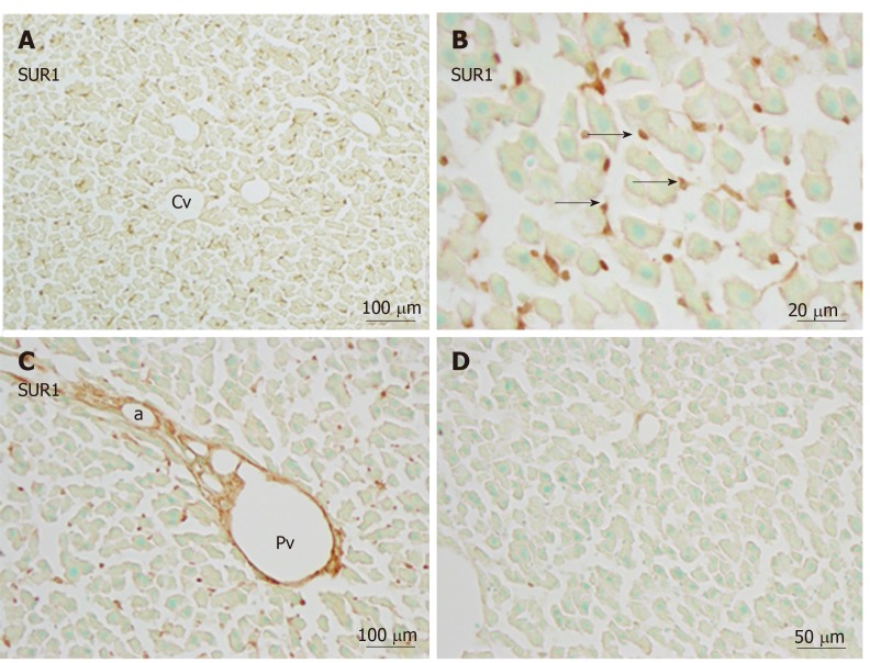
Figure 4.
Immunoreactivity with SUR1 was mainly evident in sinusoidal cells (arrows) and weaker in the cell membranes of hepatocytes. A and B: There was no such tendency toward immunoreactivity with SUR1 in the area of the central vein vs the border area; C: In the portal area, endothelial cells of the blood vessels showed intense immunoreactivity with SUR1; D: Absorption negative control section devoid of staining (no more than background) after incubation with cognate antigen peptide. a: Artery, Cv: Central vein; Pv: Portal vein. Bars: 100 µm (A and C), 20 µm (B), 50 µm (D).

