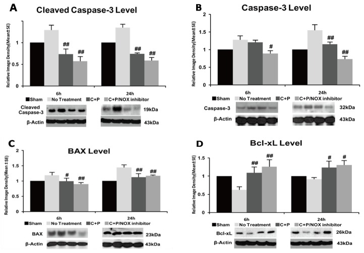
Figure 5.
Cleaved and uncleaved caspase-3, Bax, and Bcl-XL protein expression, and Western blotting in control treatment, C+P treatment, and C+P/NOX inhibitor treatment. Brain tissue containing the dorsolateral striatum and frontoparietal cortex were processed and used to detect protein levels (mean ± SE). (A) Cleaved caspase-3 protein was significantly reduced by both C+P and C+P/NOX inhibitor treatment cohorts at 6 and 24 h. Cleaved caspase-3 level at 6 h: no treatment 1.3 ± 0.1, C+P 0.7 ± 0.1, C+P/NOX inhibitor 0.6 ± 0.1; cleaved caspase-3 level at 24 h: no treatment 1.3 ± 0.1, C+P 0.7 ± 0.0, C+P/NOX inhibitor 0.6 ± 0.1. (B) Uncleaved caspase-3 protein was significantly reduced only in the C+P/NOX inhibitor cohort at 6 h. At 24 h, both C+P and C+P/NOX inhibitor cohorts resulted in decreased caspase-3; caspase-3 protein expression in the C+P/NOX inhibitor cohort was further decreased in comparison to the C+P cohort. Caspase-3 level at 6 h: no treatment 1.3 ± 0.1, C+P 1.2 ± 0.1, C+P/NOX inhibitor 0.9 ± 0.0; PKC level at 24 h: no treatment 1.5 ± 0.2, C+P 1.2 ± 0.1, C+P/NOX inhibitor 0.7 ± 0.1. (C) At 6 h, Bax protein expression was reduced in both C+P and C+P/NOX inhibitor treatment cohorts. At 24 h, both C+P and C+P/NOX inhibitor treatment cohorts exhibited a significant decrease in Bax protein expression. Bax level at 6 h: no treatment 1.2 ± 0.1, C+P 1.0 ± 0.1, C+P/NOX inhibitor 0.9 ± 0.0; Bax level at 24 h: no treatment 1.4 ± 0.1, C+P 1.1 ± 0.1, C+P/NOX inhibitor 1.2 ± 0.0. (D) At 6 h, both C+P and C+P/NOX inhibitor treatment cohorts resulted in a significant increase in Bcl-XL protein expression. At 24 h, both C+P and C+P/NOX inhibitor treatment groups also produced a significant increase. Bcl-XL level at 6 h: no treatment 0.6 ± 0.1, C+P 1.1 ± 0.2, C+P/NOX inhibitor 1.3 ± 0.1; Bcl-XL level at 24 h: no treatment 0.9 ± 0.0, C+P 1.2 ± 0.2, C+P/NOX inhibitor 1.3 ± 0.1 (# p < 0.05, ## p < 0.01).

