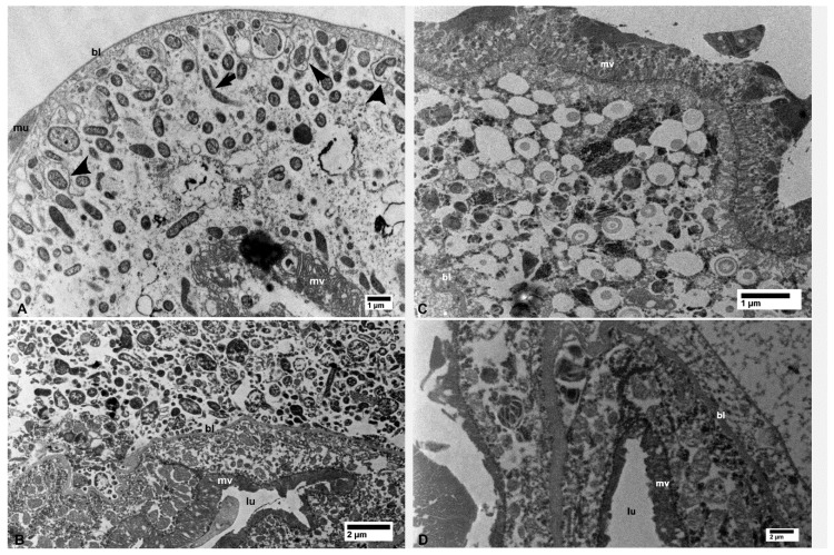
Figure 2.
Bacterial cells in the midgut (A) and filter chamber (B) of CLas-infected D. citri. A. In the midgut, bacterial cells are within the vacuolated areas either free in the cytoplasm or located between the infoldings of the basal plasma membranes (arrowheads); the arrow indicates an apparently dividing bacterial cell. (B) Large aggregates of bacterial cells outside the midgut basal lamina (bl), within the filter chamber (top half of the figure, above the basal lamina). No similar bacterial cells were found in the uninfected midgut (C) and filter chamber (D). Abbreviations: bl—basal lamina; lu—midgut lumen; mu—muscles; mv—microvilli. (A: osmicated tissue; B–D: non-osmicated).

