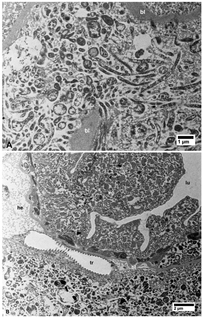
Figure 3.
(A) Details of bacterial aggregates outside the midgut within the filter chamber of CLas-infected D. citri. (B) Aggregates of bacteria inside epithelial cells (ep) of the hindgut and outside the hindgut basal lamina (bl) in CLas-infected D. citri. Abbreviations—bl, basal lamina; ep—epithelial cells of the hindgut; he—hemocele; lu—hindgut lumen; tr—trachea. (A and B: non-osmicated tissues).

