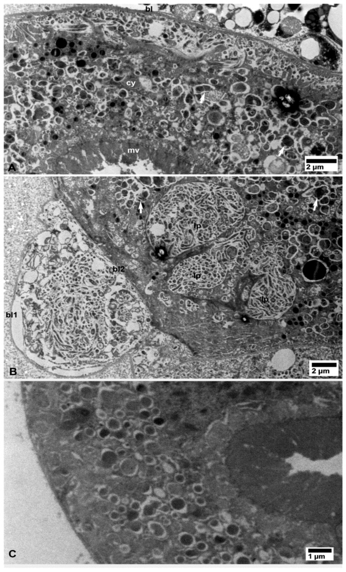
Figure 4.
Bacterial cells in the Malpighian tubules of CLas-infected (A,B) D. citri. (A) Aggregates of bacterial cells under the basal lamina (bl) and in small pockets in the cytoplasm (cy). (B) Bacterial cell aggregations between the two layers of the basal lamina (bl1, bl2) as well as within large pockets (lp) in the cytoplasm. Arrows indicate putative bacterial multiplication sites(also shown below in other organs at higher magnifications). No bacterial cells aggregates were found in similar tissues from healthy control psyllids (C). Other abbreviations: mv—microvilli; bl—basal lamina; and cy—cytoplasm. (A–C: non-osmicated tissues).

