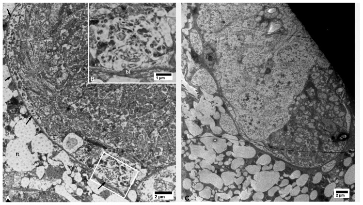
Figure 9.
Small aggregates of bacterial cells (arrows) under the basal lamina (bl) of neural tissues in CLas-exposed D. citri (A,B); the boxed area in panel (A) is shown at a higher magnification in panel (B). No bacterial cells were found in similar tissues from healthy control psyllids (C); ft—fat tissue.

