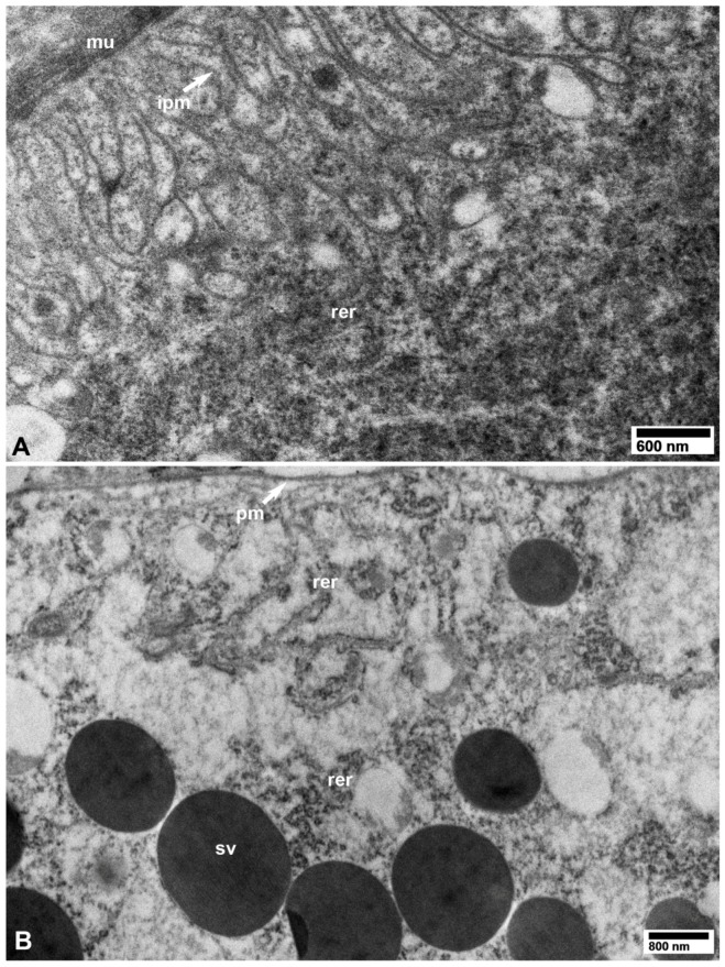
Figure 11.
TEM images from healthy control D. citri adults (unexposed to CLas). (A) Basal part of a midgut epithelial cell, showing surrounding muscle fibers (mu), infoldings of the basal plasma membrane (ipm), and rough endoplasmic reticulum (rer). (B) Part of a secretory cell in the salivary gland showing plasma membrane (pm), secretory vesicles (sv), and rough endoplasmic reticulum (rer). (A,B: osmicated tissues).

