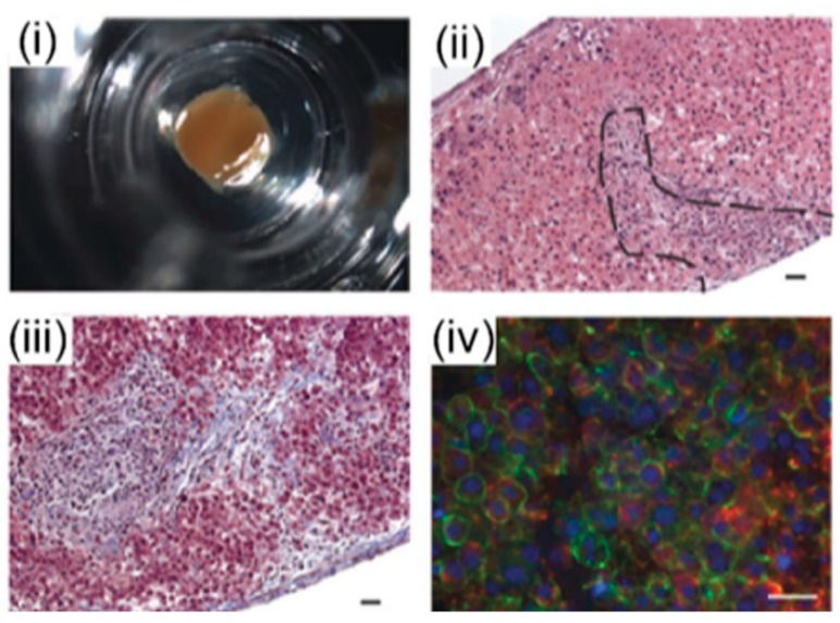
Figure 6.
Organovo’s mini liver tissue: (i) A macroscopic image of liver tissue housed in a 24-well transwell, (ii) Hematoxylin and eosin (HE) staining of a tissue cross-section, (iii) extracellular matrix (ECM) deposition assessed by Masson’s trichrome staining, and (iv) Ιimmunohistochemistry (IHC) staining of the parenchymal compartment for E-cadherin (green) and albumin (red) [71].

