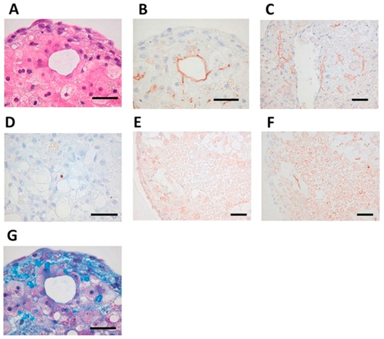
Figure 8.
Self-organization in bio-printed human liver tissues. (A) Hematoxylin and eosin stain (HE) staining shows structure of bio-printed liver tissue on day 50. (B) Immunostaining with the MRP2 antibody detected bile acid transporters (day 50). (C) Immunostaining with, cluster of differentiation 31 (CD31) antibody detected blood vessel-like and sinusoid-like structures (day 14). (D) Terminal deoxynucleotidyl transferase dUTP nick end labeling (TUNEL) staining detected little apoptosis (day 60). (E) Immunostaining with the OAT2/8 antibody detected drug uptake transporters (day 44). (F) Immunostaining with MRP2 antibody showed tissue distribution (day 44). (G) Masson’s trichrome staining shows collagen accumulation (day 50). Black bars represent 50 μm [82].

