Figure 9.
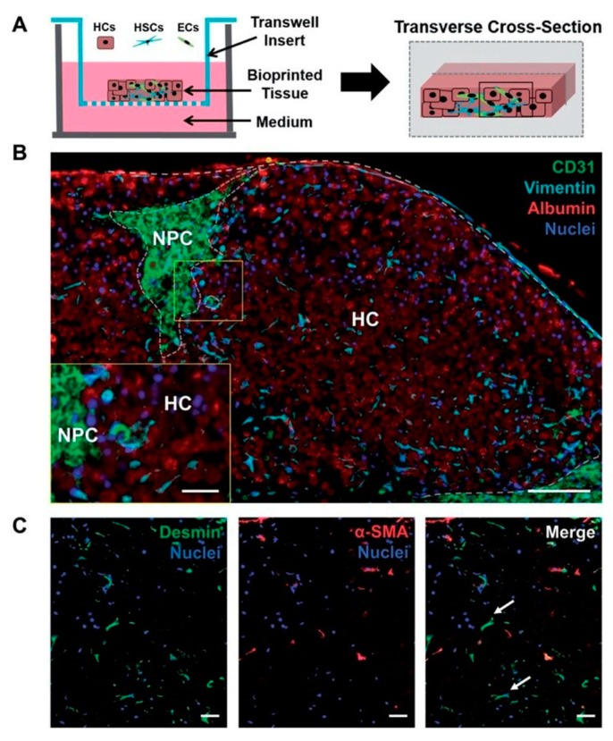
3D bioprinted tissue exhibits a compartmentalized architecture and maintains hepatic stellate cells in a quiescent-like phenotype. (A) Illustration of a transverse cross-section of bioprinted tissue on a transwell insert comprising hepatocytes (HCs) and compartmentalized endothelial cells (ECs) and hepatic stellate cells (HSCs). (B) The organization of non-parenchymal cells (NPCs) is depicted with CD31 and vimentin staining to mark ECs and HSCs, respectively. Albumin is used to denote the hepatocellular compartment (HC). Scale bar = 100 μm, inset scale bar = 25 μm. (C) HSC activation status was examined using desmin (generic marker) and Alpha-smooth muscle actin (α-SMA) (activation marker). Quiescent HSCs are denoted with white arrows. Scale bar = 50 μm [95].

