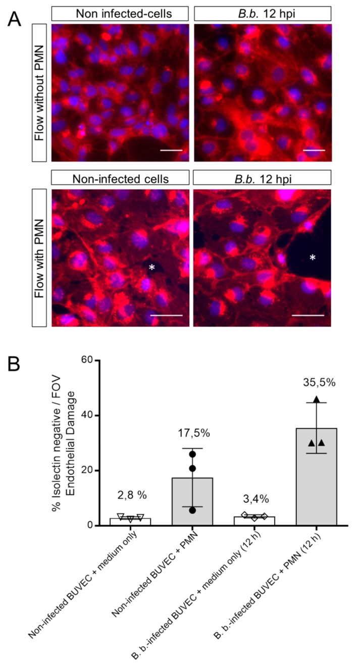
Figure 3.
PMN induce damage on B. besnoiti-infected BUVEC under physiological flow conditions. PMN or medium alone were perfused at a constant shear stress of 1 dyn/cm2 over B. besnoiti-infected and non-infected BUVEC at 12 h.p.i. After 5 min of perfusion, cell layers were fixed and stained with DAPI for nuclei and with Alexa Fluor 594-conjugated isolectin-IB4 that predominantly binds endothelium and observed under fluorescence microscopy. Endothelial damage is calculated dividing the isolectin-negative surface (white asterisks, A, representative images) by the total surface of the field of view. Column graph represents results of the percentage of endothelial damage after analyzing five random pictures from three different BUVEC isolates per each experimental condition (B). FOV = Field of view. Number over the bars indicates the mean % and error bars ± SD.

