Figure 4.
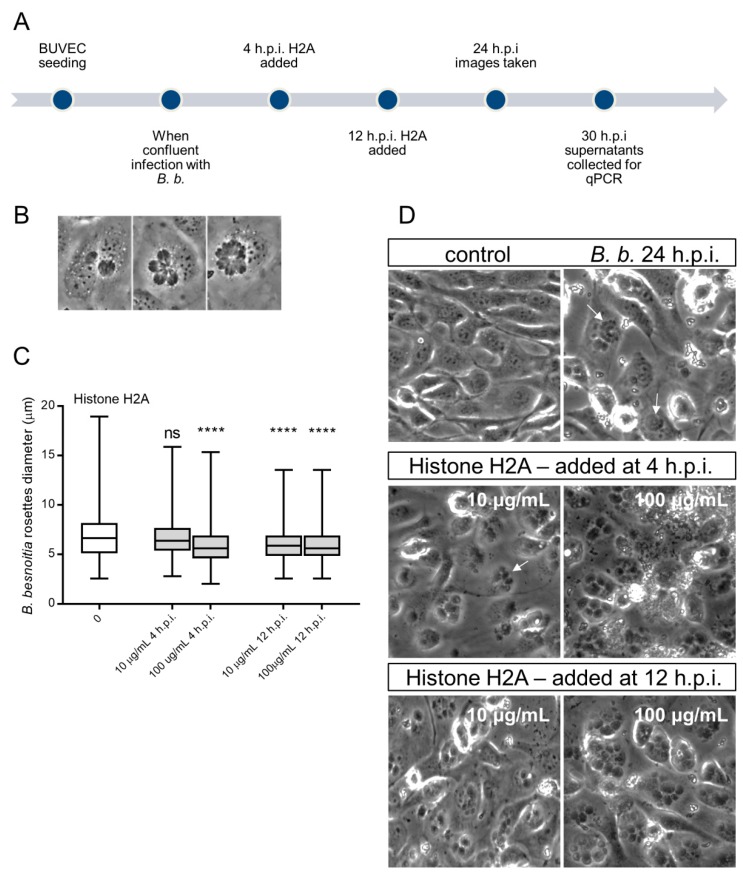
Histone 2A (H2A) treatment of B. besnoiti-infected BUVEC reduces B. besnoiti parasitophorous vacuole (PV) diameter. BUVEC (three different isolates, N = 3) were treated with H2A at 10 or 100 µg/mL at 4 and 12 h.p.i. (for experimental procedure refer to Figure 4A). At 24 h.p.i. experimental conditions were documented by five randomly taken images using a phase contrast microscope (B,D). (B) Shows the typical development of B. besnoiti rosettes within 24 h of infection (non-synchronous tachyzoite division leads to the formation of 2-mers to 16-mers). Here, the diameter of each B. besnoiti PV was measured (n = 825) and plotted as box and whiskers plot (C), line at median, bars indicating maximum and minimum values. Statistical significance, N = 3 (ns = non significant, **** p < 0.0001) was determined by Kruskal-Wallis test followed by a Dunn´s post-test comparing experimental versus control condition at 4 and 12 h.p.i. In (D) a representative image of each experimental condition is shown, white arrows indicate B. besnoiti rosettes inside host BUVEC cell.

