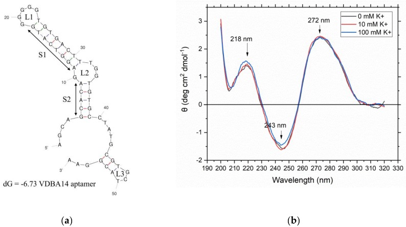
Figure 2.
Investigation of the structural properties of the used VDBA14 aptamer. (a) Predicted secondary structure of the VDBA14 aptamer using mFold. Stem structures are marked as “S”, whereas loop structures are marked as “L”. The calculated initial free energy was dG = −6.73. (b) Circular dichroism spectrum of the VBDA14 aptamer in the presence of three different binding buffer conditions with increasing potassium concentrations (0–100 mM K+).

