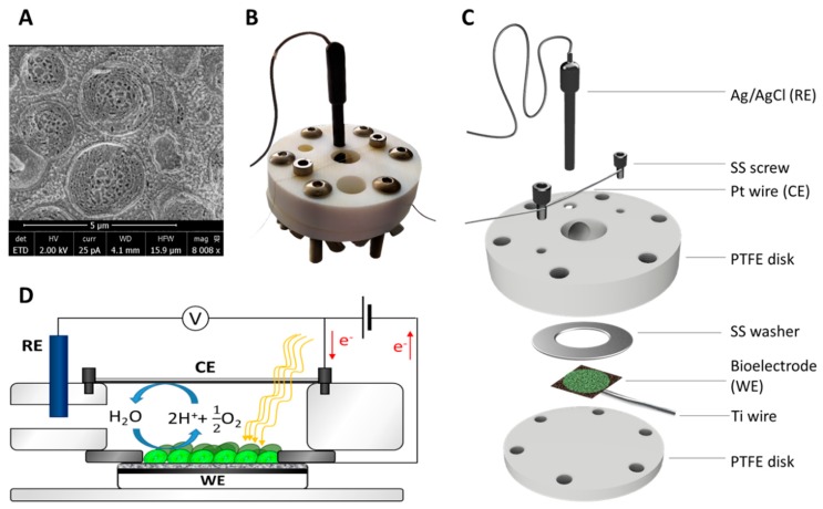
Figure 2.
(A) Cryo-SEM image of Synechocystis cells, (B) picture, (C) semi-exploded view, and (D) electrochemical diagram of the setup used for the experiments. The bioelectrode (working electrode, WE) was clamped using two PTFE disks which also held the platinum wire (counter electrode, CE) and the Ag/AgCl reference electrode (RE). The stainless-steel washer ensured electrical connection between the bioelectrode and the titanium wire.

