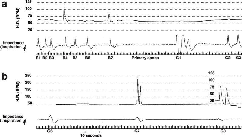
Figure 1.
Recordings of heart rate (H.R.) by ECG and respiratory activity by thoracic impedance in an infant classified as a SIDS case. (a) Breaths (B) 1 through 7 show a slowing of respiratory rate (i.e. progressively longer apnea) in a background of severe bradycardia (Normal HR = ~150 bpm). As hypoxia becomes more severe respiratory activity ceases altogether (primary hypoxic apnea). Three gasps (G1–3) then emerge, the respiratory component of “autoresuscitation”. (b) Terminal gasps (6 through 8) in the same record are shown – note that gasping has not succeeded in elevating H.R. or re-establishing eupnea; i.e. failed autoresuscitation. Reproduced from Sridhar et al., 2003.

