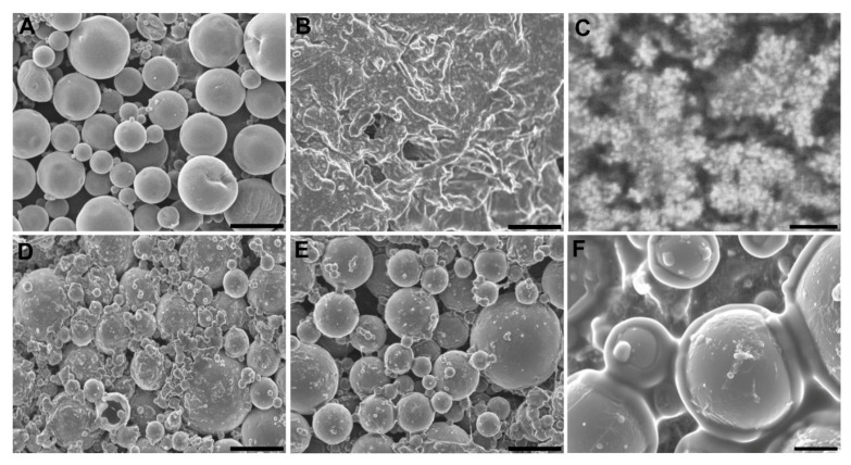
Figure 1.
SEM images (A) microspheres, (B) hydrogel, (C) hydrogel high magnification detail, (D) core system freshly prepared, (E) internal structure of the core system after 4 weeks incubation in water at 37 °C and 25 rpm, and (F) high magnification detail of Figure 1E. Scale bars: (A–E) 100 µm, (C) 1 µm, (F) 20 µm.

