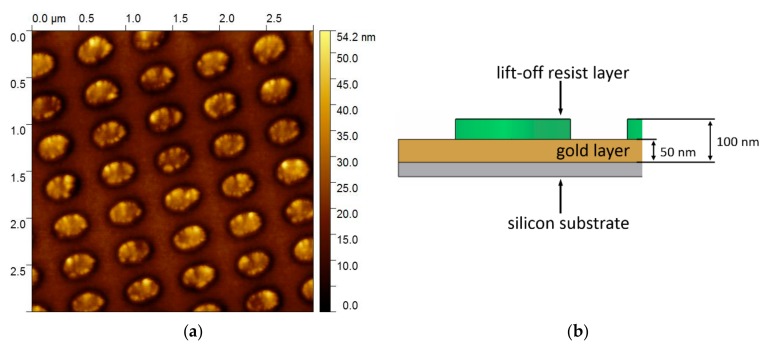
Figure 7.
Scanning force microscopy (SFM) image of the substrate after the tape transfer process and schematic sketch of the layer thicknesses. (a) The remaining elliptical pillars can be observed by SFM imaging. The height of the imaged structure is indicated by the color bar at the right of the SFM image. (b) Schematic sketch of the cross section of the sample indicating the layer thicknesses.

