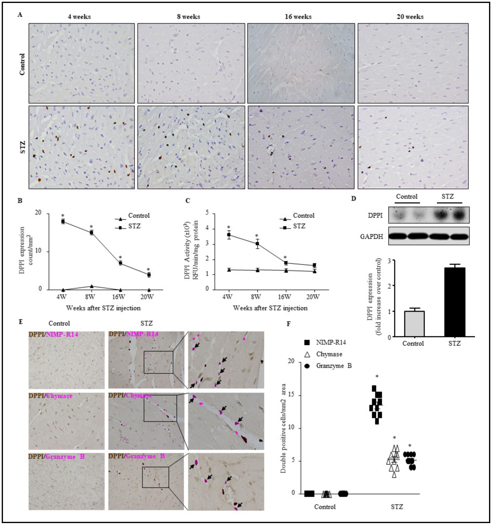
Fig. 1.

STZ treatment increases leukocyte infiltration, DPPI expression and activation. LV sections from animals treated with citrate buffer or STZ for 4–20 weeks were assessed for anti-DPPI immunostaining (400X magnification with scale bars 50 μm) (A) and quatification (B), DPPI activity as determined by specific fluorogenic substrates (C), and DPPI immunoblot analysis (D). Double immunistaining with DPPI and NIMP-R14 (neutrophil), chymase (mast cells), or granzyme B (cytotoxic T cells) antibodies (400X magnification with scale bars 50 μm) (E) and quantification (F), respectively. n=5 for each group *=p< 0.05 vs control. One-way ANOVA followed by the Tukey post hoc test was used to compare multiple groups. Two-way ANOVA and subsequent Tukey test were performed to compare groups with different time points.
