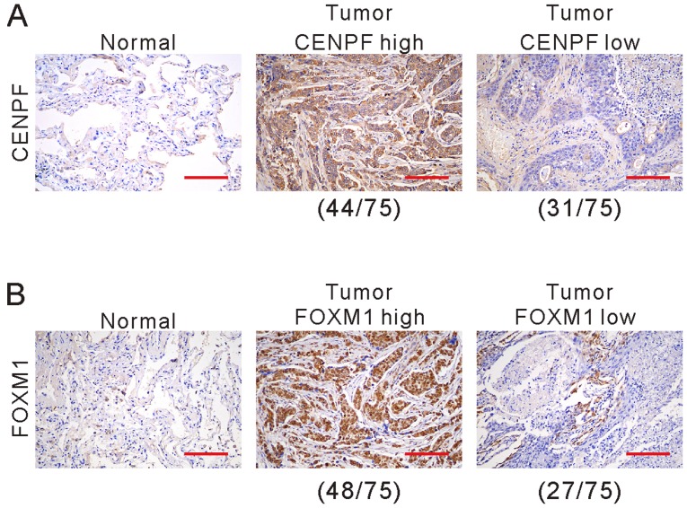
Figure 3.
Immunohistochemical staining of CENPF (A) and FOXM1 (B) in NSCLC and non-cancerous tissues. The positive staining for CENPF and FOXM1 is brown in cytoplasm and nucleus. Nucleus is blue in hematoxylin counterstaining. Scale bars, 100 µm. NSCLC, non-small cell lung cancer; CENPF, centromere protein F; FOXM1, Forkhead box M1.

