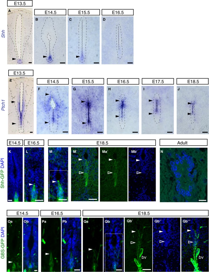
Figure 6.

Shh activity persists after decline in Shh transcripts during mouse spinal cord development. (A–D) In situ hybridisation showing Shh mRNA expression in the floor plate (arrowheads) at the indicated stages (E13.5: 14 sections, n = 3 embryos; E14.5: 15 sections, n = 5 embryos; E15.5: 58 sections/explants, n = 9 embryos; E16.5: 38 sections/explants, n = 9 embryos). From E17.5 onwards, Shh mRNA expression was no longer detected in the spinal cord (E17.5: 38 sections/explants, n = 8 embryos; E18.5: 18 sections, n = 3 embryos; 24‐ and 40‐week‐old: 17 sections, n = 3 adult mice; data not shown). (E–J) Ptch1 mRNA expression at the indicated stages (E13.5: 27 sections, n = 5 embryos; E14.5: 26 sections/explants, n = 5 embryos; E15.5: 43 sections/explants, n = 6 embryos; E16.5: 49 sections/explants, n = 8 embryos; E17.5: 40 sections/explants, n = 8 embryos; E18.5: 16 sections, n = 3 embryos). Note Ptch1 is excluded from the floor plate and detected in ventral half of the ventricular layer (between arrowheads) and persists in the forming central canal, but was no longer detected in adult (40‐week‐old: 24 sections/explants, n = 3 mice; data not shown). (K–Mb’’’) Immunofluorescence for GFP in Shh‐GFP embryos was detected in the floor plate during development (arrowheads) including dissociating ventral cell population at later stages (outlined white arrowhead): E14.5: 17 sections, n = 1 embryo; E16.5: 17 sections, n = 1 embryo; E18.5: 18 sections, n = 1 embryo, (M’‐Mb’ single z‐plane confocal images of region shown in white dashed‐line square in M and more ventral domain) (n) but not in adult central canal (9–10 week adult (88 sections, n = 3 mice); (Oa–Qb’’’). Immunofluorescence for GFP (arrowheads) in GBS‐GFP embryos revealed positive cells in the ventral region at indicated stages (E14.5: 47 sections, n = 3 embryos; E16.5: 43 sections, n = 3 embryos; E18.5: 33 sections, n = 3 embryo) (Qb’–Qb’’’ single z‐plane confocal images of region shown in white dashed‐line square in Qb and more ventral domain). K–M’, Mb’, n, Ob, Pb, Qb qb’’, Qb’’’ nuclei are stained with DAPI (blue). bv, auto‐fluorescent blood vessel. Scale bars: 40 μm, except n: 10 μm.
