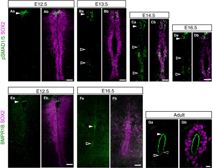
Figure 7.

Expanding BMP signalling and BMPR1B expression during mouse spinal cord development. (Aa–Bb) Immunofluorescence for phospho‐SMAD1/5 (green) detected in a dorsal cell population at E12.5 (16 sections, n = 2 embryos) and E13.5 (16 sections, n = 3 embryos) (full arrowheads), phospho‐SMAD1/5 was also detected in the ventral ventricular layer at E13.5 (outlined arrowhead); (Ca–Db). At E14.5 (36 sections, n = 3 embryos) and E16.5 (14 sections, n = 2 embryos), phospho‐SMAD1/5 was detected in dorsal (full arrowheads) and ventral regions of the ventricular layer (outlined arrowheads). (Ea–Fb) BMPR1B (green) was enriched dorsally at E12.5 (10 sections, n = 1 embryo) (full arrowhead) and was detected dorsally and apically in ventricle abutting cells (between arrowheads) at E16.5 (23 sections, n = 3 embryos). (Ga,Gb) Pronounced apical localisation of BMPR1B in adult central canal (10‐ to 11‐week‐old mice: 22 sections, n = 2 mice), which was more weakly detected ventrally (dashed outline arrowhead). Progenitor cells within the ventricular layer are labelled with SOX2 (magenta). Scale bars: 50 μm, except Adult: 10 μm.
