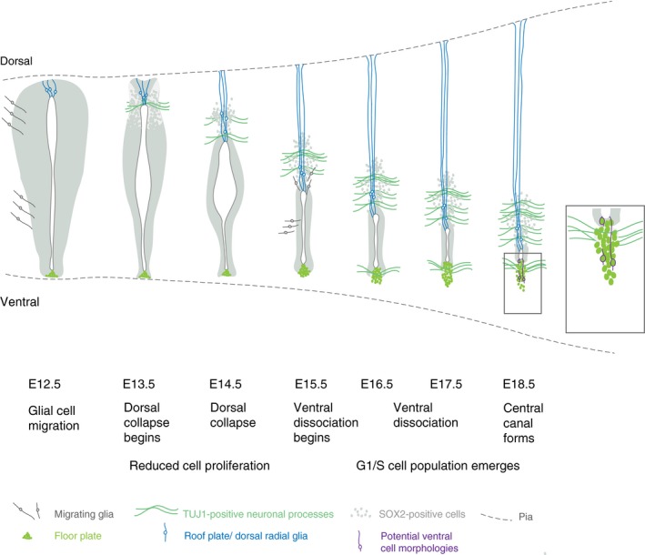
Figure 8.

Summary of steps contributing to spinal cord ventricular layer attrition. Schematic of key changes in cell arrangement and cell cycle in the spinal cord ventricular layer at daily intervals leading up to the formation of central canal. SOX2‐positive ventricular cells are indicated in grey (note that floor plate cells are also SOX2‐positive, but distinguished here in bright green, and SOX2 is also expressed in astrocytes at later stages). Glial cell migration from the ventricular layer begins prior to dorsal collapse (Deneen et al. 2006; Stolt et al. 2003) and distinct glia cell populations continue to emerge from the ependymal cell population forming the central canal, including dorsal and lateral/ventral astrocytes from E15.5 (Hochstim et al. 2008; Li et al. 2018). Dorsal collapse is accompanied by elongation of roof plate cells, which give rise to dorsal radial glia (in blue) (Xing et al. 2018; Shinozuka et al. 2019), appearance of the dorsal commissure (TUJ1‐positive cell processes) and reduced proliferation of ventricular layer cells (this study). These events are followed by dissociation of ventral‐most floor plate cell group (the nuclei of these cells are not included in the lining of forming central canal, but some of these cells may retain a contact with its apical surface, potential ventral cell morphologies are indicated in purple), accompanied by appearance of the ventral commissure (TUJ1‐positive cell processes) and an increase in the percentage of cells in G1/S phase of the cell cycle (this study).
