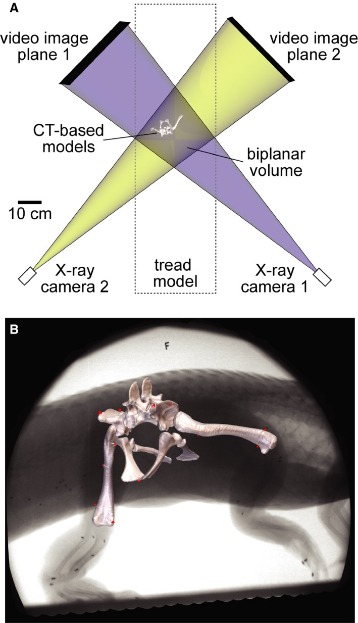
Figure 1.

XROMM setup reconstructed as a maya scene. (A) Top view of treadmill representing the two X‐ray systems as pairs of virtual X‐ray cameras and video image planes. The blue and yellow beams overlap in the biplanar volume, allowing bone and soft tissue models to be animated based on the rigid body transformations of marker clusters. (B) Models of the pelvis and femora registered to X‐ray Video S1 showing the locations of surgically implanted conical markers (red).
