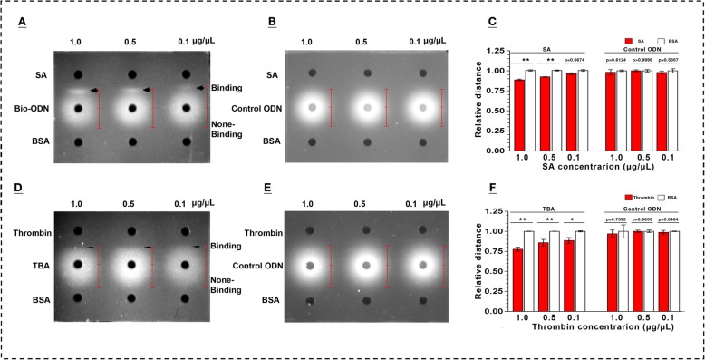
Fig. 2.
Characterization of oligodeoxynucleotides (ODN)-target binding by double-diffusion in mini-gels. Diffusion profiles of ODN modified with biotin (Bio-ODN) (a) and without biotin (control ODN) (b) during diffusion toward target streptavidin (SA) and non-target bovine serum albumin (BSA), respectively. Quantitative analysis of diffusion distance of ODN from the central point of different loading wells to their diffusion edge (c & f). Diffusion profiles of thrombin aptamer (TBA) (d) and non-aptamer ODN (control ODN) (e) when they diffused toward target thrombin and non-target BSA, respectively. Indication of binding: shortened diffusion distance due to stagnation of diffusion caused by the formation of binding complexes at the interface of the double-diffusion. Arrow heads in a: appearance of precipitation line of binding complexes at the interface of the double-diffusion. Thin arrow heads in d: interface of the double-diffusion of aptamer and its target; Data represent mean ± SEM. *P < 0.05, **P < 0.01, set control = 1.0. GelRed as the DNA indicator

